
Release date:2026-01-15 Browse:
Cryo-Electron Microscopy (Cryo-EM) is an electron microscopy technique used to image biological macromolecules, organelles, and even whole cells at cryogenic temperatures. Samples are rapidly frozen to preserve them in a near-native, hydrated state, thereby avoiding structural artifacts introduced by chemical fixation or staining. Its key advantage lies in its ability to determine high-resolution three-dimensional structures of proteins and complexes without the need for crystallization, achieving atomic-level resolution while capturing dynamic processes in different conformational states. Cryo-EM plays a vital role in life science research, widely used for elucidating the structures of membrane proteins, giant complexes, and viruses, thereby advancing drug design, disease mechanism studies, and the development of novel biomaterials.
Why You Need This Service (Using Proteins as an Example)
• Your material is available in limited quantities.
• The material is difficult to crystallize.
• You are investigating atomic-level details of the molecule.
• The sample is a large biomolecular complex.
• You aim to observe the true, native-state structure.
The Longlight provides professional, one-stop Cryo-EM solutions, covering the entire process from sample preparation and imaging to advanced data modeling. Clients can choose our comprehensive recommended service package, individual modular services, or engage in customized protocol development to suit diverse research project needs. Leveraging our advanced technological platform and efficient workflows, we facilitate the rapid acquisition of high-resolution structural information, driving high-quality research output and innovative breakthroughs.
From Sample Prep to Data Analysis
1. Cryo-EM Single Particle Analysis (Cryo-EM SPA)
• Resolution achievable: 2-3 Å
• Deliverables: 3D map, atomic model, validation report
• Suitable for: Proteins, antibody-antigen complexes, membrane proteins
2. Cryo-Electron Tomography (Cryo-ET)
• Resolution achievable: 2-3 Å
• Deliverables: 3D map, atomic model, validation report
• Suitable for: Proteins, antibody-antigen complexes, membrane proteins
3. Negative Staining Characterization
• Rapid assessment of sample homogeneity and integrity
• Commonly used for preliminary screening
4. Data Processing & Modeling Service
• Automated reconstruction, resolution assessment, atomic model building
• Personalized analysis solutions
5. Customized Solutions
• Technical consultation, method development
• Long-term collaboration & joint R&D
We offer an integrated workflow covering sample preparation, image acquisition, and structure determination, with advantages in fast turnaround, exceptional quality, and efficient cross-team/cross-platform collaboration.
Featured Cryo-EM Services
1. Cryo-EM Single Particle Analysis (SPA) Service
Utilizing our Cryo-EM platform and image processing pipeline to accurately analyze biomacromolecules in a near-native state, obtaining high-quality 3D density maps and atomic models.
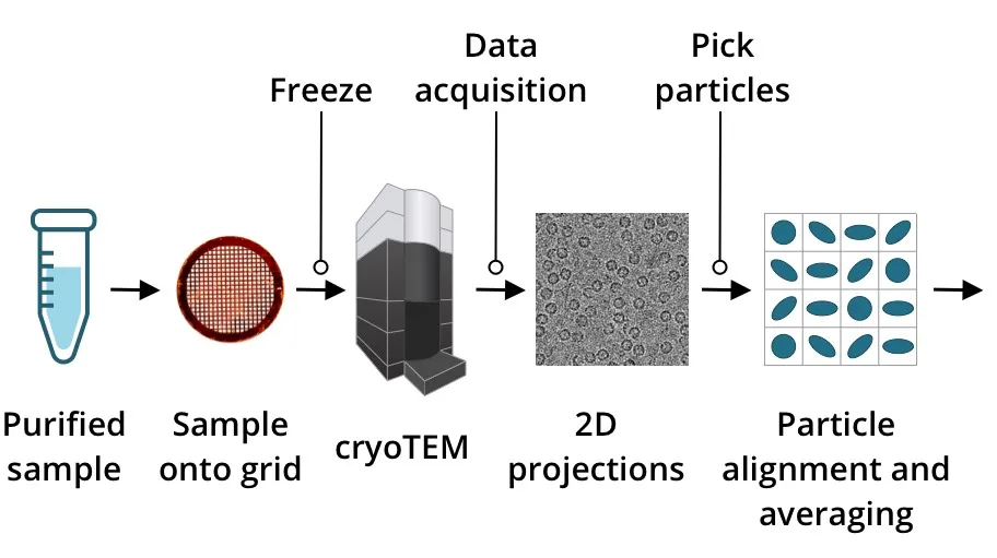
Workflow
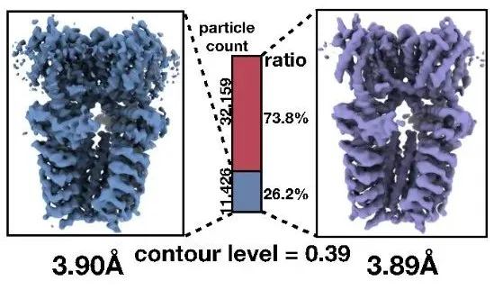
Output
Application
• Membrane proteins & receptor complexes
• Viral structure & entry mechanism
• Virus-antibody interactions
•Neuroscience & disease mechanisms
2. Cryo-Electron Tomography (Cryo-ET) Analysis Service
Based on our Cryo-EM platform, employing tilt-series acquisition and 3D reconstruction to study biomolecular structures in their native cellular context and analyze interactions within cells.
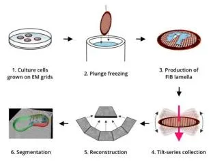
Workflow
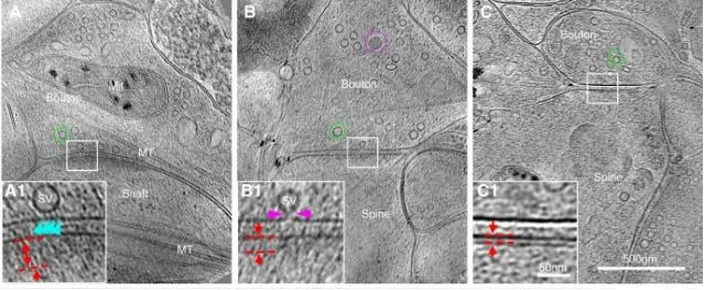
Output
Application
• Dynamic biological processes & in situ pharmacology
• Cell biology & organelle studies
• Viral replication mechanisms & host interactions
• Virus-host interface studies
• Neurodegenerative disease mechanisms
• Plant science & synthetic biology
• High-resolution bio/nanostructure imaging
FAQ
• How does Cryo-EM differ from other structural biology techniques?
Cryo-Electron Microscopy offers several advantages compared to other structural biology techniques like X-ray crystallography and NMR spectroscopy. Cryo-EM enables direct imaging of molecules in their intrinsic, biologically relevant state without the need for crystallization or heavy chemical staining. Unlike X-ray crystallography, Cryo-EM can also image larger and more complex molecules, making it particularly useful for studying viruses and large macromolecular complexes.
• What is the complete service workflow for a Cryo-EM project?
Project consultation → NDA signing → Service agreement confirmation;
Sample reception and quality check;
Negative stain pre-screening → Determination of vitrification conditions;
Grid preparation and preliminary screening → Condition optimization;
Large-scale, automated data collection;
Data processing and 3D reconstruction;
Resolution assessment and structure determination;
Project delivery and reporting.
• What are the specific deliverables from a Cryo-EM service?
Raw microscope images (micrographs);
Processing results (2D class averages, 3D map);
FSC curve and local resolution map;
Atomic model (if requested);
Complete technical service report (including methodology, results presentation, and technical parameters).
• Can you assist with preparing research papers or patent applications?
We can assist clients in organizing figures, drafting methodological descriptions, and, with client authorization, participate in the publication of results.
Ordering Process
Consultation → Sample preparation & shipping → Proposal evaluation → Data acquisition → Data processing → Report delivery.


 粤公网安备44030002008509号